-
Viewpoint on 'IONS'
Viewpoint on 'Scientific Literacy'
- Proudly sponsored by
-


-
A Wind Tunnel for Quantum Physics

Simulating quantum phenomena on todays computers can be extremely challenging. Yet, just like the wind tunnel changed the trajectory of modern aviation, new specially built quantum simulators may soon guide the design of tailor-made quantum materials.
-
Spooky Light
Many are afraid of darkness, but who gets spooked by light? Light has now been used to condition rats to fear, teaching us about fear memory formation.
-
The Number of Life

1/137 give or take: the value of the fine-structure constant. Our Universe, our life and everything we know depend on this number. What would the consequences be, if this were to change?
Volume 17 Story 1 - 16/4/2012

 Due to their ability to see through materials, X-rays are routinely used by physicians to diagnose broken bones and also by airport security staff to scan passengers luggage. This very quality — the generally weak interaction of X-rays with matter — that makes them so useful, however, also makes them extremely difficult to handle — much more difficult, in fact, than most other kinds of electromagnetic waves such as visible light. When building X-ray lasers, the challenges are particularly evident: unlike the readily available visible-light lasers, X-ray lasers are typically built around gigantic particle accelerators. Researchers led by Nina Rohringer, from the Lawrence Livermore National Laboratory in California (USA), have now built a novel type of laser that generates better-defined X-rays. This may prove advantageous for studying biological and crystalline structures.
There is more to light than meets the eye. In fact, visible light is but a tiny portion of the electromagnetic spectrum which also includes X-rays. All electromagnetic waves are characterized by their wavelength: the shorter the wavelength, the more powerful and aggressive the wave. X-rays have extremely short wavelengths, between 0.01 and 10 nanometers. This makes them very useful. For example, their ability to see through materials is very convenient for medical imaging. Furthermore, since the smallest details that can be seen in microscopy are proportional to the wavelength of the electromagnetic radiation, X-rays are frequently the tool of choice when it comes to imaging extremely tiny crystalline and biological structures. Thus, they have an impact on fundamental and applied research in physics, chemistry, crystallography, medicine, biology and many other fields. However, this superlative resolving power comes at a price: X-rays are so aggressive that they end up damaging the samples under study. Indeed, because X-rays are ionizing radiation, unlike the light used in optical lasers, DVDs, and laser pointers, the tendency is for the X-ray photons to alter the sample.
Technological innovation over the past decades has proven the potential of lasers: most light sources — including the Sun, fire, lightning, and light bulbs — emit light at a whole spectrum of wavelengths. Lasers, by contrast, emit light only at an extremely specific wavelength. This specificity turns out to be a great asset for ultra-fast and ultra-precise applications. Therefore, visible-light and infrared lasers are the technological backbone for our optical communications systems, many sensing applications, as well as a long list of scientific studies. Of course, X-ray lasers are far less common than this kind of lasers. Nonetheless, they may well become very useful for imaging proteins and other biological structures with unprecedented speed and accuracy.
X-ray lasers are far more complex than the laser pointers we are used to. "An X-ray laser facility," Rohringer explains, "is typically connected to a gigantic particle accelerator: an electron beam is accelerated to nearly the speed of light and sent through an array of strong magnets with alternating polarizations. This facility is called a Free Electron Laser (FEL). The interaction of these extremely fast electrons with the strong magnets creates the X-ray laser beam." However, the X-ray photons the particles of light produced by the FELs are not all identical to one other, which means that the quality of the laser is far from perfect.
Rohringers X-ray laser produces extremely aggressive, high-quality X-rays: "we built the shortest-wavelength X-ray laser that works according to the same principles as conventional visible-light lasers," Rohringer says. In the case of conventional lasers, photons are produced when the excited electrons of an atom jump back to their ground state. In order to squeeze X-rays out of an atom, however, Rohringers group needed to use another X-ray laser. "We used the laser beam created by the Linac Coherent Light Source (LCLS) at the Stanford Linear Accelerator Center (SLAC) in California (USA) as the base for our experiments. This X-ray laser beam was focused onto a target of neon atoms, thereby ionizing them. Spontaneous emission of the ionized neon atoms then led to stimulated emission by other atoms."
The higher quality of the X-ray laser beam compared to other currently available technologies is the key feature of Rohringers laser, according to Linda Young [1,2] from Argonne National Laboratory in Illinois (USA): "The achievement is important because an X-ray laser based upon atomic transitions can possess wavelength stability and temporal coherence considerably superior to current FELs. The superior wavelength stability has been demonstrated in Rohringer's paper. However, the improved temporal coherence has, to date, only been inferred from simulation."
"X-ray lasers represent a new scientific playground," concludes Young. In fact, these new lasers receive so much attention because their high-energy photons can be used to work with atoms and molecules in completely new ways: "X-ray lasers provide the ability to probe matter simultaneously on atomic length- and time-scales, and the tunability of such X-ray lasers will provide access to elemental and chemical specificity; this will mean that these lasers will become important tools for research, as one strives to make technology smaller, cheaper, and faster. A primary application for X-ray lasers has been the promise of single biomolecule imaging at atomic resolution without the need for crystallization, currently a bottleneck in structural biology and drug design."
[1] L. Young et al., Femtosecond electronic response of atoms to ultra-intense X-rays, Nature 466, 56-61 (2010).
[2] T. E. Glover et al., Controlling X-rays with light, Nat. Phys. 6, 69-74 (2010).

Towards Better X-ray Lasers
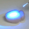
Red, green and blue lasers all produce extremely good-quality light and therefore play a key role in everyday life. X-ray lasers, in comparison, are still in their infancy. Building on the principles of visible-light lasers, however, it is possible to produce better X-rays.Artistic representation of an X-ray laser. X-rays interact with the electrons of the atom, which leads to the emission of another X-ray photon. Credit: Illustration by Gregory M. Stewart, SLAC National Accelerator Laboratory.
There is more to light than meets the eye. In fact, visible light is but a tiny portion of the electromagnetic spectrum which also includes X-rays. All electromagnetic waves are characterized by their wavelength: the shorter the wavelength, the more powerful and aggressive the wave. X-rays have extremely short wavelengths, between 0.01 and 10 nanometers. This makes them very useful. For example, their ability to see through materials is very convenient for medical imaging. Furthermore, since the smallest details that can be seen in microscopy are proportional to the wavelength of the electromagnetic radiation, X-rays are frequently the tool of choice when it comes to imaging extremely tiny crystalline and biological structures. Thus, they have an impact on fundamental and applied research in physics, chemistry, crystallography, medicine, biology and many other fields. However, this superlative resolving power comes at a price: X-rays are so aggressive that they end up damaging the samples under study. Indeed, because X-rays are ionizing radiation, unlike the light used in optical lasers, DVDs, and laser pointers, the tendency is for the X-ray photons to alter the sample.
Technological innovation over the past decades has proven the potential of lasers: most light sources — including the Sun, fire, lightning, and light bulbs — emit light at a whole spectrum of wavelengths. Lasers, by contrast, emit light only at an extremely specific wavelength. This specificity turns out to be a great asset for ultra-fast and ultra-precise applications. Therefore, visible-light and infrared lasers are the technological backbone for our optical communications systems, many sensing applications, as well as a long list of scientific studies. Of course, X-ray lasers are far less common than this kind of lasers. Nonetheless, they may well become very useful for imaging proteins and other biological structures with unprecedented speed and accuracy.
X-ray lasers are far more complex than the laser pointers we are used to. "An X-ray laser facility," Rohringer explains, "is typically connected to a gigantic particle accelerator: an electron beam is accelerated to nearly the speed of light and sent through an array of strong magnets with alternating polarizations. This facility is called a Free Electron Laser (FEL). The interaction of these extremely fast electrons with the strong magnets creates the X-ray laser beam." However, the X-ray photons the particles of light produced by the FELs are not all identical to one other, which means that the quality of the laser is far from perfect.
Rohringers X-ray laser produces extremely aggressive, high-quality X-rays: "we built the shortest-wavelength X-ray laser that works according to the same principles as conventional visible-light lasers," Rohringer says. In the case of conventional lasers, photons are produced when the excited electrons of an atom jump back to their ground state. In order to squeeze X-rays out of an atom, however, Rohringers group needed to use another X-ray laser. "We used the laser beam created by the Linac Coherent Light Source (LCLS) at the Stanford Linear Accelerator Center (SLAC) in California (USA) as the base for our experiments. This X-ray laser beam was focused onto a target of neon atoms, thereby ionizing them. Spontaneous emission of the ionized neon atoms then led to stimulated emission by other atoms."
The higher quality of the X-ray laser beam compared to other currently available technologies is the key feature of Rohringers laser, according to Linda Young [1,2] from Argonne National Laboratory in Illinois (USA): "The achievement is important because an X-ray laser based upon atomic transitions can possess wavelength stability and temporal coherence considerably superior to current FELs. The superior wavelength stability has been demonstrated in Rohringer's paper. However, the improved temporal coherence has, to date, only been inferred from simulation."
"X-ray lasers represent a new scientific playground," concludes Young. In fact, these new lasers receive so much attention because their high-energy photons can be used to work with atoms and molecules in completely new ways: "X-ray lasers provide the ability to probe matter simultaneously on atomic length- and time-scales, and the tunability of such X-ray lasers will provide access to elemental and chemical specificity; this will mean that these lasers will become important tools for research, as one strives to make technology smaller, cheaper, and faster. A primary application for X-ray lasers has been the promise of single biomolecule imaging at atomic resolution without the need for crystallization, currently a bottleneck in structural biology and drug design."
[1] L. Young et al., Femtosecond electronic response of atoms to ultra-intense X-rays, Nature 466, 56-61 (2010).
[2] T. E. Glover et al., Controlling X-rays with light, Nat. Phys. 6, 69-74 (2010).
Armand Niederberger
2012 © Optics & Photonics Focus
AN is a SU2P Research Fellow working on quantum networks at Stanford University, California, USA.
Nina Rohringer, Duncan Ryan, Richard A. London, Michael Purvis, Felicie Albert, James Dunn, John D. Bozek, Christoph Bostedt, Alexander Graf, Randal Hill, Stefan P. Hau-Riege & Jorge J. Rocca, Atomic inner-shell X-ray laser at 1.46 nanometres pumped by an X-ray free-electron laser, Nature (2012) 481, 488-491 (link).