-
Viewpoint on 'IONS'
Viewpoint on 'Scientific Literacy'
- Proudly sponsored by
-


-
Flat Light from a Flat Diamond
The possibility to polarize light in optical fibers comes to establish graphene as a likely key player in the future of optical technologies; a new application emerges for this material that rocked the scientific world due to its fascinating properties.
-
Journey to the Center of the Earth
What happens when we put an electric insulator, such as iron oxide, under pressure and temperature conditions as extreme as those of the Earths interior?
-
Holography Goes Beyond 3D
Holography is an inherently 3D technology that now supports full color. It could, therefore, provide the ultimate 3D experience, with images and movies appearing identical to the real world and without the need for 3D glasses.
Neurons, Freeze!
Neurons are the building block of the arguably most complex structure of the Universe: the human brain. Recent experiments show innovative ways to shock-freeze neurons while they are communicating with each other.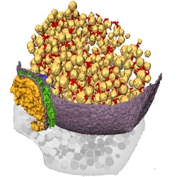
Reconstruction of a synapse. A 3D representation of the presynaptic neuron. The yellow spheres are the vesicles filled with neurotransmitter. The different types of filaments are in red and blue, the membranes of the presynaptic and postsynaptic neuron are in purple, and the synapse is in green. The postsynaptic neuron cotent is represented in dark yellow.
Organisms are made of cells. In complex organisms like humans specialized cells take over specific functions and work together with other cells. Neurons are the cells constituting nerve fibers. They are responsible for transport and treatment of sensorial and motor information in our body. Neurons need to communicate information between each other and they do so in different ways. One example is the reaction between visual stimulus and muscular contraction, such as an escape reaction: "theres a hungry lion, run away!"
Neurons are also fundamental building blocks in our brain and thus are ultimately responsible for all forms of imagination and reasoning. The space where two neurons exchange information is called synapse. The sending presynaptic neuron releases neurotransmitter molecules that pass through the synaptic cleft and dock to the receptors on the receiving postsynaptic neuron.
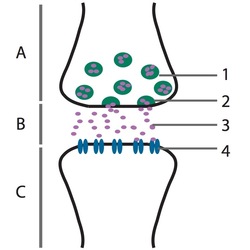
How neurons communicate. In the presynaptic neuron (A) vesicles filled with neurotransmitter molecules (1) fuse with the presynaptic neuron membrane (2) to release the neurotransmitter molecules (3). These pass through the synaptic cleft (B), also called active zone, and bind to the corresponding receptors (4) at the postsynaptic neuron (C).
What researchers usually do in order to study the presynaptic neuron structure is to freeze the whole synaptic region with many neurons and then image the sample using a powerful microscope. Electron microscopes, in particular, provide high resolution images well beyond the capabilities of optical microscopes. By combining images taken from different angles with a technique known as electron tomography it is even possible to obtain a 3D image of the sample.
Unfortunately, there is a big problem that arises when freezing the sample: water forms ice crystals and thus alters the neuronal structure. Worse still, ice crystals may even destroy the cellular structure or the components under investigation. In fact, water significantly expands during the formation of ice, which is why ice floats on water and also the reason why one should never place a completely full and sealed glass bottle into the refrigerator. The research team in Munich was able to come up with a solution to this problem which will allow a more accurate study of the neuronal structure. "The problems associated with ice crystals can be avoided by shock-freezing the sample," Lučić explains. His team froze the synaptic region using a novel technique called vitrification.
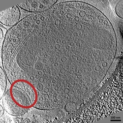
A shock-frozen synapse. This image shows a synapse that passed though the vitrification process and was obtained by electron microscopy. It is one of the images used for the 3D reconstruction. In the red circle is a synapse, on the top is the presynaptic neuron with its neurotransmitter vesicles and on the bottom is the postsynaptic neuron.
In this way, Lučić's team was able to immobilize presynaptic neurons without any alterations due to the formation of ice, which brought them closer to the actual physiological configuration. They studied, in particular, the filaments connecting vesicles containing the neurotransmitter. "These filaments," Lučić explains, "are involved in the regulation of the synaptic release. Whenever a neuron has to release its vesicles, these filaments regulate their mobilization and facilitate the release of the neurotransmitter. Also, when there is no need to release the neurotransmitter, these filaments limits vesicle dispersion."
"Lučić's experiment," Serge Marty from École Normale Supérieure in Paris, France, says, "enables us to distinguish real structures from artifacts." Conventional methods did allow for studies of the structure of the presynaptic neuron, but they could not rule out errors originating from ice crystals. "Other methods that are used to immobilize the presynaptic neuron," Marty continues, "also use freezing but they also dehydrate the sample causing some damage to the neuron's structure." Marty therefore sees this new method as an interesting complement to conventional methods that observe synapses in neurons without the need of the same protective substances.
"At this point," Lučić adds, "we are very interested in the kind of molecules that constitute the different types of filaments. Also we would like to understand the relationship between the different types of filaments as well as their exact functions." Both scientists are confident that the understanding of many complex physiological processes will benefit from studies of vesicular dynamics. This understanding could even shed new light on the ever elusive and ever captivating question of how we actually think.
María-Belén Pérez Ramírez
2010 © Optics & Photonics Focus
MBPR is a research student working in neurophysiology at the National Autonomous University of Mexico (UNAM, México, D.F.).

Rubén Fernández-Busnadiego, Benoît Zuber, Ulrike Elisabeth Maurer, Marek Cyrklaff, Wolfgang Baumeister & Vladan Lučić, Quantitative analysis of the native presynaptic cytomatrix by cryoelectron tomography, The Journal of Cell Biology (2009) 188, 145156 (link).