-
Viewpoint on 'IONS'
Viewpoint on 'Scientific Literacy'
- Proudly sponsored by
-

Neurons, Freeze!
Optics & Photonics Focus
Volume 9 Story 1 - 13/4/2010
Optics & Photonics Focus
Volume 9 Story 1 - 13/4/2010
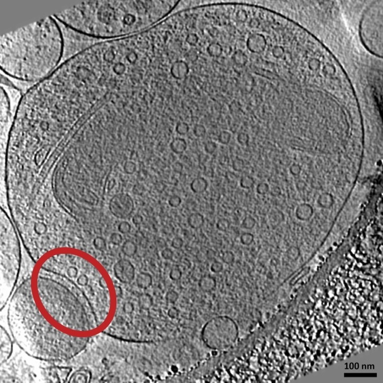
A shock-frozen synapse
This image shows a synapse that passed though the vitrification process and was obtained by electron microscopy. It is one of the images used for the 3D reconstruction. In the red circle is a synapse, on the top is the presynaptic neuron with its neurotransmitter vesicles and on the bottom is the postsynaptic neuron.
[read full story]